Radiation department
- Home
- Radiology Department
greeting
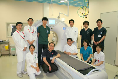
The Department of Radiology is in charge of the diagnostic imaging center (general radiography, CT / MR), endoscopy center, RI / radiotherapy center (nuclear medicine / linac / cyberknife), and IVR center (angiography). In each department, we work in cooperation with department staff including Registered Nurse Radiological Technologist is becoming more and more important in the ever-advancing medical care.
Under these circumstances, we will fulfill our responsibilities based on the department's philosophy and code of conduct.
Radiology Department Chief Hidekazu Tanaka
Radiation Department Philosophy
- We will practice team medical care in cooperation with other occupations.
- We provide high-quality images with high diagnostic value and advanced radiotherapy.
- We will endeavor to use radiation equipment safely and reduce the exposure dose.
Radiation Department Code of Conduct
- Try to give a kind and easy-to-understand explanation.
- We strive for medical safety and prevent accidents.
- Participate in academic societies, study sessions, and seminars to acquire the latest knowledge and skills.
- We support all 24-hour radiological examinations (excluding RI and radiation therapy).
- We will carry out the inspection even if it is not reserved on the day.
- We will strive to reduce the waiting time for patient examinations, the waiting time for multiple radiological examinations, and the waiting time for image data (CD, film) output.
- We will also actively accept requested inspections through the Regional Cooperation Office.
- any will continue to study with the goal of acquiring various certifications and entering conference presentations.
Diagnostic Imaging Center
We perform general radiography, contrast examination, bone mineral density examination, X-ray CT, and MRI.
General shooting
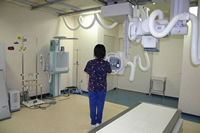
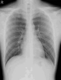
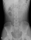
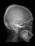
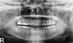
General radiography is a general term for X-ray radiography that does not use contrast media, such as radiography of the head, spine, chest, mammary glands, abdomen, limbs, and Surgery Inflammation, pain, bone deformities / fractures, gas status, presence of stones, etc. will be photographed.
Contrast examination
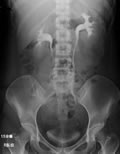
IVP (Excretory Renal Meng Urethrography) Examination
Lower limb vein examination
The examination time is about 40 minutes and there are dietary restrictions.
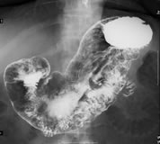
Stomach / esophagus / duodenal examination
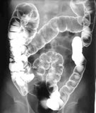
Large intestine examination (enema examination)
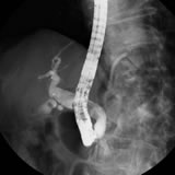
Endoscopic retrograde cholangiopancreatography
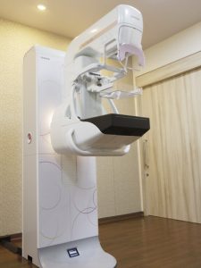
Mammography
It is an excellent test for detecting breast cancer.
Because the breast is a soft tissue, it cannot be taken clearly with a normal X-ray machine. Therefore, by using an X-ray machine dedicated to the breast, it is possible to capture "small calcifications" and "lumps" that lead to the detection of breast cancer.
In addition, as a detailed examination of "lumps", a biopsy (a needle is pierced and a "lump" is aspirated to examine cells) is performed with a mammotome device.
A female technician is in charge of photography and mammothome examination. The shooting time is 20 to 30 minutes, and the mammothome biopsy is about 1 to 2 hours.
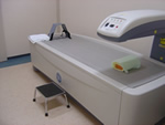
Quantitative bone mineral test (bone density test)
CT (Computed Tomography) inspection
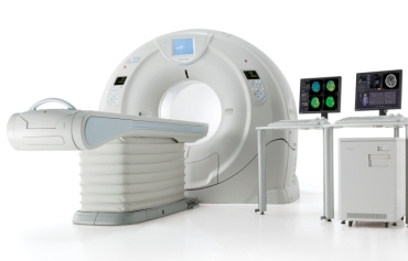
CT laboratory (320-row multi-CT)
- If metal is attached to the part to be photographed, it may interfere with the image. You will be asked to change into an inspection garment if necessary.
- If you are pregnant or may become pregnant, please let us know.
(Basically, inspection is not possible.)
Contrast Enhanced CT (Contrast Enhanced CT)
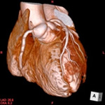
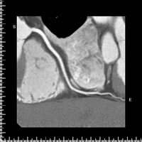
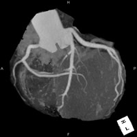
Coronary CT (cardiac CT)
- Since contrast media have side effects, please read the "Contrast Agent Manual and Consent Form" in advance. If you do not have data on renal function (serum creatinine level), please collect blood in advance. In that case, please wait until the result is obtained. Please note.
- Please do not eat for 3 hours before the test (water and tea are fine).
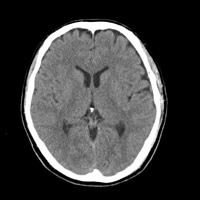
Head CT
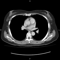
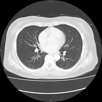
Chest CT
This is done when mediastinal disease of the lung field is suspected.
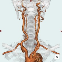
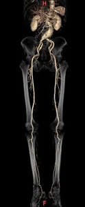
CT angiography (angiography) image
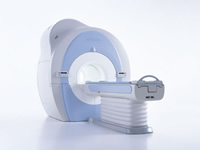
MRI (Magnetic Resonance Imaging) examination
MRI device (1.5 Tesla)
- There will be a noise during the inspection.
- People who are not good at narrow spaces may not be able to inspect.
To cooperate with patients
- Metals (hearing aids, hairpins, glasses, watches, etc.) are not allowed.
- If you are using a pacemaker, please let us know.
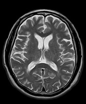
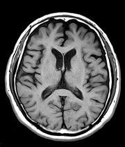
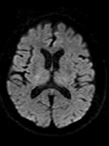
Brain MRI
Abdominal MRCP (Magnetic Resonance Cholangio Pancreatograhy)
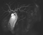
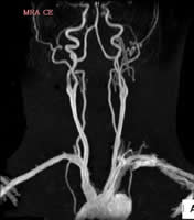
Contrast-enhanced MRI CE MRI (Contrast Enhanced MRI)
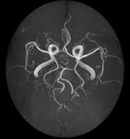
MRA (MR Angio)
IVR Center
IVR Center (interventional radiology)
We perform angiography, catheter treatment, and endovascular surgery. Collectively, these are called vascular IVR.
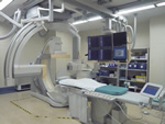
Angiography device
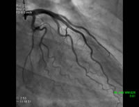
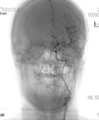
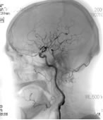
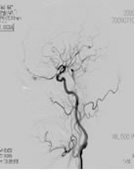
The X-ray tube rotates in multiple directions, and high-speed continuous imaging is performed in the direction in which the blood vessel you want to observe can be seen clearly. Angiography of the head, heart, chest and abdomen, upper and lower limbs, etc. can be performed.
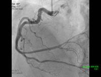
Cardiac catheterization is a test in which a catheter is inserted into a heart chamber or blood vessel under fluoroscopy, and a contrast medium is used to detect the condition of the heart or abnormalities in blood vessels.
Cerebrovascular examinations are mainly taken in two directions, front and side, and then a 3D image is created by rotation photography and observed from any angle. This is a test to check for aneurysms and bleeding.
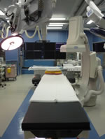
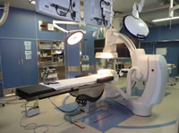
Hybrid room
Blood vesselsIt is a room equipped with imaging equipment and also has an operating room function. Specifically, vascular Surgery, Cardiovascular Surgery in Stent-graft insertion, intracerebral vascular therapy, emergency trauma etc., in the normal intravascular catheterization, cases or to consider switching to surgery for conditions, catheter We mainly deal with cases where treatment and surgery are performed at the same time.
When performing transcatheter aortic valve indwelling (TAVI), depending on the situation, there are cases where intervention is switched to emergency thoracotomy, but even in such cases, the patient can be treated on the spot without moving. It is a facility that can be done. In this room, each clinical department doctors, Registered Nurse, Radiological Technologist, Clinical Engineer while the mutual cooperation as a team, do the catheter business.
<Team example>
EVAR team: vascular Surgery physician, Anesthesiology physicians, Registered Nurse, Radiological Technologist, Clinical Engineer
TEVAR team: Cardiovascular Surgery surgeons, Anesthesiology physicians, Registered Nurse, Radiological Technologist, Clinical Engineer
Emergency Hybrid team: emergency department Surgery physicians, emergency department radiologists, Registered Nurse, Radiological Technologist
<Device>
The imaging device is an FPD biplane angiography device that allows the examination bed to be tilted, and is further combined with an operating table. It has almost the same function as our general-purpose angiography device, and can obtain much higher-definition fluoroscopy and captured images than the Surgery
It also has CT-like imaging using a 3D workstation, diagnostic support and surgery support functions such as a 3D roadmap.
<Cleanliness>
・ FED-STD-209D (US federal standard) class 1000
・ Operating table hybrid operating room system
About team medical care
Due to recent advances in medical technology, the work required of medical professionals is wide-ranging and at the same time requires specialization. Today, the role of radiologists in hospitals must be aware that they are part of a team in providing technology and provide patient-centered services. In addition, it is necessary to perform duties with sincerity and sincerity for staff of other occupations.
Hospitals consist of doctors and Registered Nurse, Pharmacist, Biomedical Laboratory Scientist, physiotherapists, Occupational Therapist, Clinical Engineer, nutritionists, radiologists, and other comedics, each of whom has an increasing role. Comedics have different basic knowledge and skills, but each requires a high degree of specialization. We must work together on medical services in the course of medical care that respects the development of new technologies and treatment methods and the QOL (Quality of life) of patients.
In addition, we are actively promoting cooperation between medical examinations (cooperation between hospitals and clinics or clinics) as stated in the hospital's philosophy of "creating better regional medical care for local people." It is possible to share expensive medical equipment such as CT / MRI / RI examinations, mammography, and bone mineral quantification examinations. Under these circumstances, the Department of Radiology will continue to practice team medical care in a broad sense so that we can provide safe and high-quality medical care centered on local patients.
RI / Radiation Therapy Center
RI (Radio Isotope) test: Nuclear medicine test
What is RI inspection?
First, a radiopharmaceutical suitable for the purpose of the test is collected by intravenous injection (or drinking) into the target organ / tissue. Since this radioactive drug contains a small amount of radioactive substances, the radiation emitted from it is detected by a dedicated camera and processed by a computer. Then, from the obtained information, we will investigate the shape, function, metabolism, etc. of the organ.
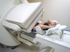
SPECT device
The SPECT device is a device that captures a small amount of radiation emitted from a radiopharmaceutical administered into the body, and a dedicated camera can collect images by bringing it close to the body.
About radiopharmaceuticals
- Radioactive drugs contain radioactive substances, but the amount is very small, the radioactivity decays quickly, and it is physiologically excreted from the body, so there is no concern about the effects on the human body due to radiation exposure.
- Radiopharmaceuticals have extremely few side effects and can be received with peace of mind.
- There are various types of radiopharmaceuticals, each of which is suitable for each test.
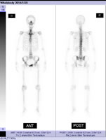
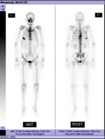
Radiopharmaceuticals that collect in the bones are administered into the body and pictures of the bones of the whole body are taken. The test will start about 3 hours after the injection. The inspection time is about 30 to 40 minutes. This test mainly checks for the presence or absence of bone metastases in the cancer.
Bone metastases appear black on the photo, but some are lacking in shape. In addition, fractures and degenerative diseases of the spine also appear black.
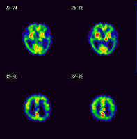
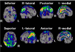
Radiopharmaceuticals (there are two types) that are taken into brain tissue from blood vessels in the brain are administered into the body to examine the blood flow status in the brain. The test will start at the same time as the injection. The inspection time is about 30 to 40 minutes. This test mainly diagnoses cerebrovascular accidents and Alzheimer's dementia.
The obtained data is analyzed by a computer to create an image as shown in the above figure. The upper left figure is a cross-sectional view of the brain, and the blood flow state is expressed in an easy-to-understand manner by displaying the cerebral blood flow distribution in color. And the figure on the upper right is called the image statistical analysis method, and by comparing it with the normal brain, you can visually see only the place where the blood flow is reduced. This image statistical analysis method is used as an auxiliary role for doctors to make a diagnosis.
Radiopharmaceuticals taken into the heart muscle from the blood vessels of the heart are administered into the body to examine the blood flow status of the heart muscle. This test involves two tests, one at rest and one at load. In the load test, either exercise load using a bicycle or drug load using a drug is performed. The inspection time is about 2 hours. This test looks at the diagnosis of ischemic myocardium, the decision of cardiac catheterization, and the recovery of blood flow after the treatment.
The obtained data is analyzed by a computer, and the heart is sliced as shown in the upper left figure to create an image as shown in the upper right figure. By displaying the blood flow distribution of the myocardium in color, the blood flow state is expressed in an easy-to-understand manner.
Through the Regional Medical Cooperation Office, we provide services for regional medical institutions to use our RI tests. After making a reservation at the regional medical cooperation room in advance, we will perform the examination at our hospital. And we will send you the test result at a later date.
In addition, there are the following items for inspection.
- Bone scintigraphy (search for bone metastases)
- Gallium scintigraphy (search for inflammatory diseases)
- Cerebral blood flow scintigraphy ECD (diagnosis of dementia)
- Cerebral blood flow scintigraphy IMP (diagnosis of cerebrovascular accident)
- Myocardial perfusion scintigraphy TF (diagnosis of ischemic myocardium)
- Myocardial sympathetic scintigraphy (diagnosis of Parkinson's disease)
Radiation therapy
Our radiotherapy is operated by a team of 2 doctors, Radiological Technologist, Registered Nurse
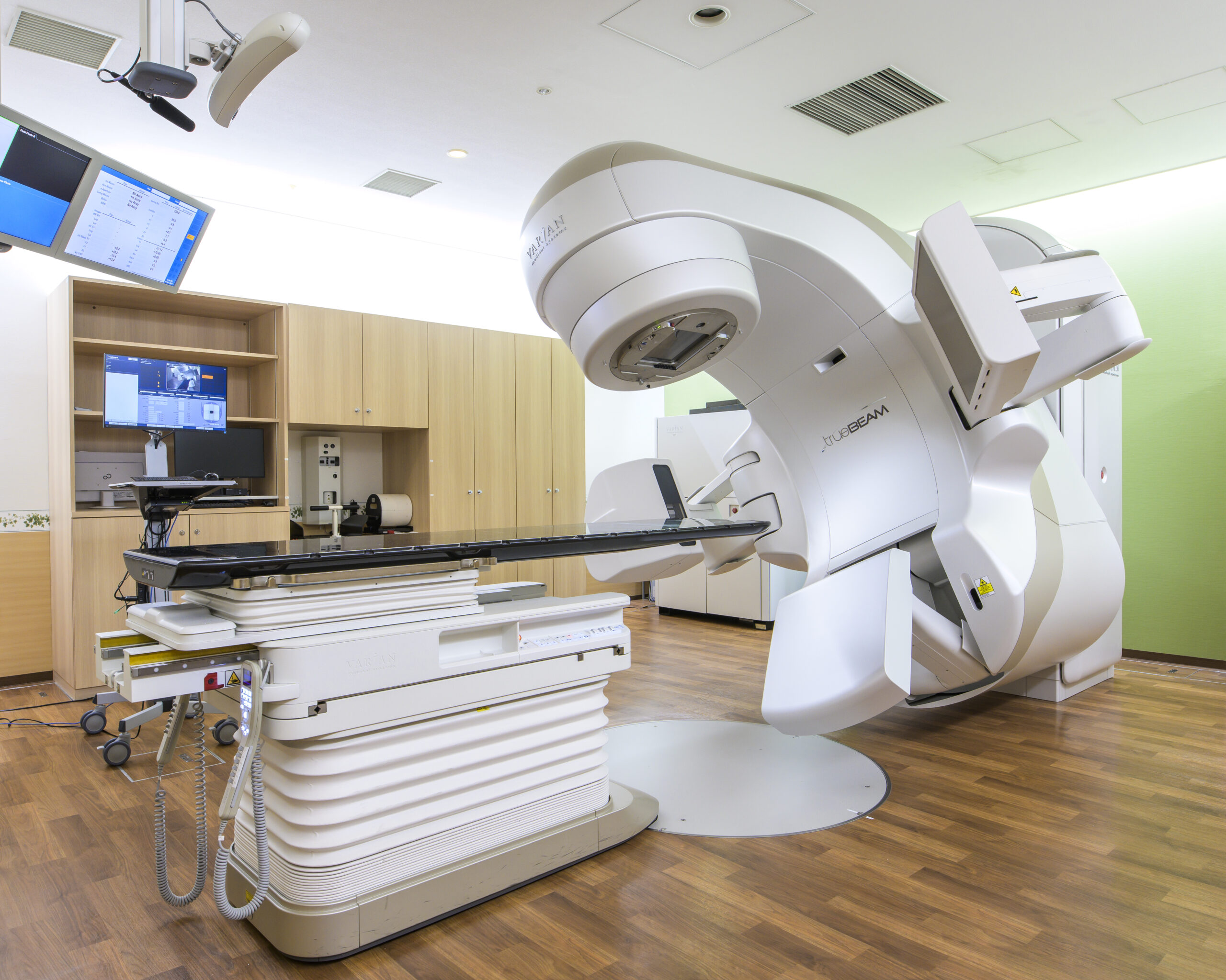
The energy of our linac device can generate a total of 8 beams of 6, 10 MV for X-rays and 5, 6, 8, 10, 12, and 14 MeV for electron beams. Energy is selected and performed by considering the size and location of the disease. In addition, since a part called a multi-leaf collimator is attached, it is possible to irradiate the beam to a shape that matches the shape and size of the tumor, so it is possible to minimize the exposure dose to normal tissue. ..
The actual flow of radiation therapy is first examined by a radiation therapist, then a CT for treatment planning is taken and the CT data is used to plan the irradiation plan. On the first day of radiation therapy, a confirmation photo called "linacgraphy" is taken, so the total time required is about 20 to 40 minutes. Once this is confirmed, mark it with an ink called skin ink. From the second day onward, the body position will be adjusted according to the mark on the first day and treatment will be performed, so the time required will be about 10 to 20 minutes. In addition, a confirmation photo may be taken again to confirm that the irradiation position has not shifted on the way.
In addition, we will provide regular medical examinations, so if you have any questions or concerns regarding radiation therapy, please feel free to ask.
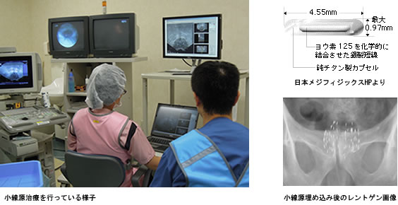
Intra-tissue irradiation performed at this hospital is a method of implanting an isotope called iodine 125 in the prostate. Iodo 125 is a nuclide that emits low-energy photons with a half-life of 59.4 days and a low energy photon of 27.4 to 35.5 keV. Is possible.
Also, since it is permanently embedded, there is no need to take it out. The procedure takes about 3 hours and is usually hospitalized for 2 days and 1 night.
Side effects include frequent stools and defecation pain, bleeding, frequent urination and micturition pain, radiation dermatitis and diarrhea. After the end of radiation therapy, the side effects diminish over time. Check with your doctor in advance about side effects. No matter how much the symptoms of prostate cancer improve, it is not always the best choice if it causes severe side effects. Also, if you have any side effects and are having a hard time, please do not overdo it and consult your doctor.
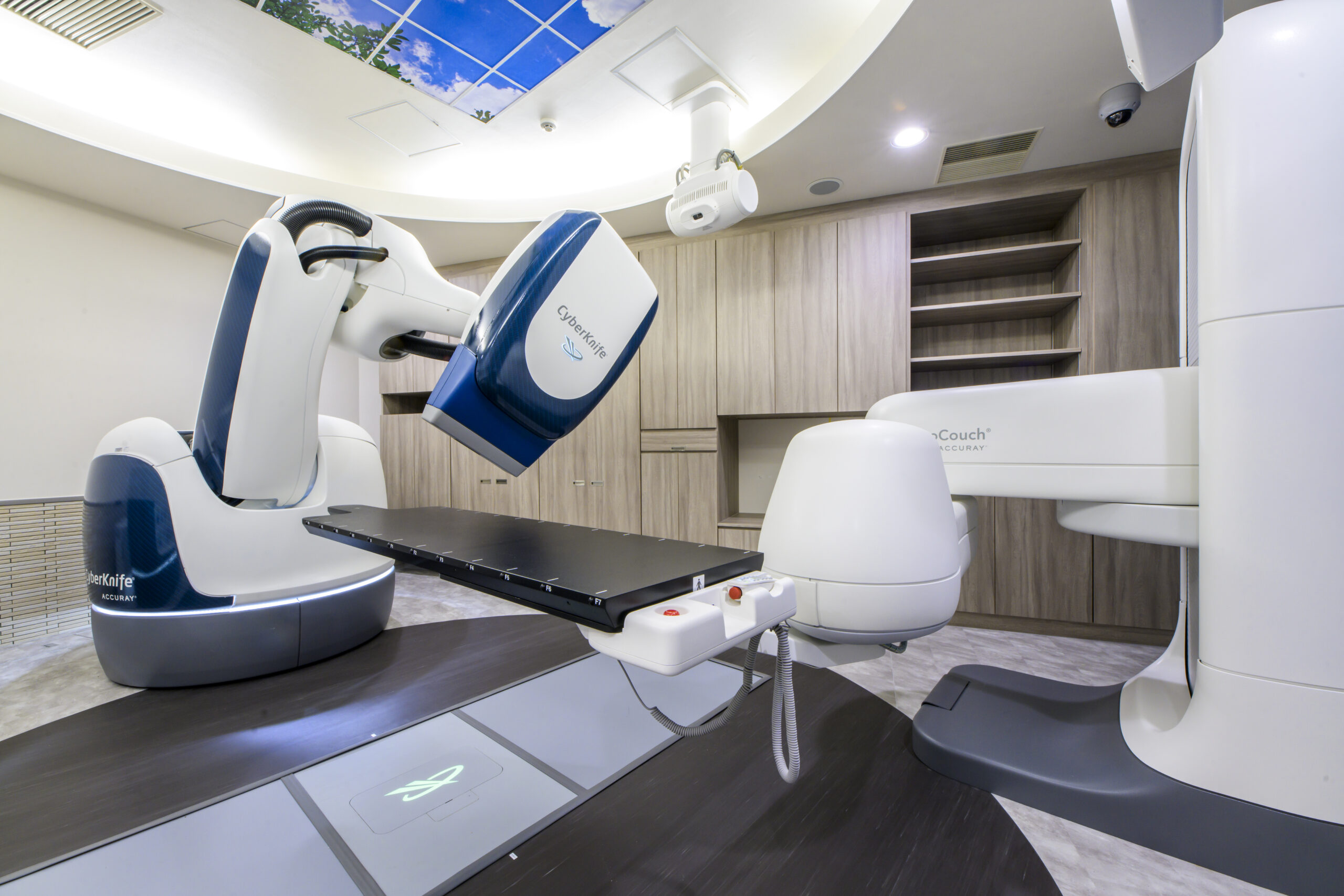
1. What is Cyberknife?
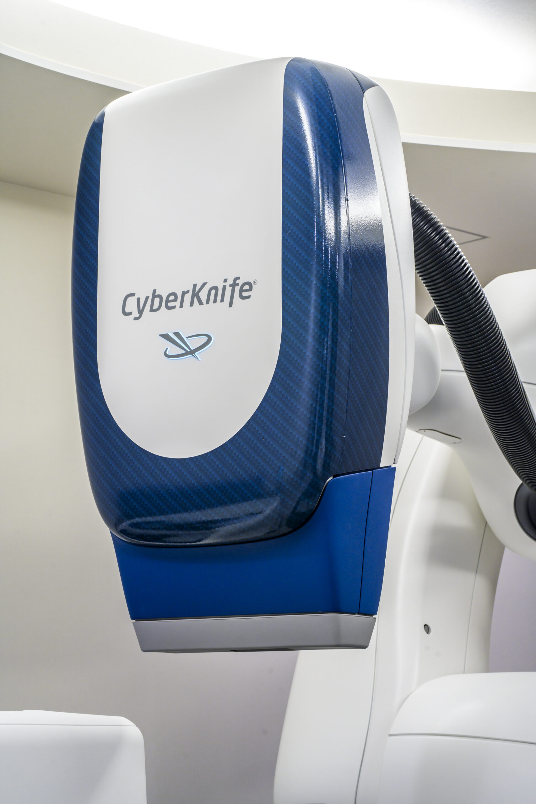
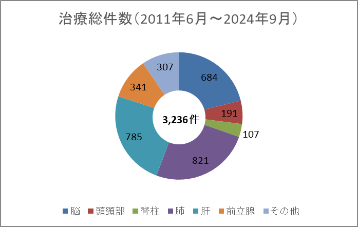
Cyberknife treatment is an epoch-making treatment method that combines a high-precision robot arm with an X-ray irradiation device. During treatment, even slight movements of the patient are detected three-dimensionally, and the deviation is immediately corrected, and the robot accurately aims at the lesion from multiple directions. It is possible to concentrate radiation only on the lesion and minimize the effect on surrounding normal tissues.
2. Comparison with Gamma Knife
a.) Fixing method
Surgery uses screws to fix the skull under local anesthesia. With Cyberknife, you simply cover it with a simple mesh-like fixture called a shell.
b.) Treatment target site
Gamma knife can only treat the inside of the skull, but Cyberknife can treat the head and neck and trunk.
3. Features of Cyberknife
a.) Compatible with various shapes and parts
The robot arm with 6 joints can irradiate from up to 1200 directions, and can handle complicated tumor shapes with an accuracy of mm or less.
b.) Automatic tracking system
During treatment, a low-energy X-ray source and a flat-mounted detector capture high-resolution images. At the same time, it is also monitored by a respiratory detector, and even if the patient moves, including those caused by breathing, the position of the tumor is automatically corrected and tracked. If it moves significantly, the irradiation will be stopped automatically, the position will be confirmed again, and the treatment will be resumed.
c.) Dose distribution
Cyberknife selects tissues that you do not want to irradiate in advance and uses a recursive treatment plan (inverse planning) that determines the irradiation direction and dose, so you can obtain an ideal dose distribution for tumors and normal tissues. You can do things. Treatment planning is done in the reverse process of conventional radiation therapy.
4. Number of treatments
It has little effect on the normal tissue around the lesion and can be irradiated with a large dose only to the lesion. Therefore, the time required for one irradiation is longer than that of conventional radiation therapy, but the period of radiation therapy is shorter, and it is possible to reduce the impact on the patient's daily life. The number of treatments is one to several times, but the number of irradiations is appropriately determined according to the type and size of the lesion.
5. Cases to be treated
Indications are one of the following:
・ Primary brain tumor (diameter 3 cm or less)
・ Metastatic brain tumor (3 cm or less in diameter, 3 or less, and general condition can withstand this treatment)
・ Localized head and neck tumor (size that can set the irradiation range of this treatment)
・ Cerebral arteriovenous malformation
・ Local recurrence after whole-brain irradiation (diameter 3 cm or less)
From December 2013, Toshiba's 80-row CT Aquilion PRIME started operation in the Radiation Therapy Planning Office.
In current radiation therapy, CT is taken for treatment planning in all cases. The position and range of the tumor are identified based on the CT image, and the irradiation range and irradiation amount are determined. With Aquilion PRIME, 80-row helical scan enables imaging in a short time, which shortens the patient's breath holding and enables clear images. In addition, since it is equipped with the latest image processing function, it is possible to diagnose and plan with smooth and less blurred images.
The entrance to the CT has also become wider, making it possible to take pictures using the fixtures used for radiation therapy, and the device has less oppressive feeling.
With Aquilion PRIME, it has become possible to continuously photograph an area of 4 cm, and the movement of the tumor can be visually observed. Pinpoint treatment using Cyberknife introduced in our hospital requires irradiation that accurately grasps the movement of the tumor and enhances concentration, but pinpoint treatment is possible more than ever. Therefore, effective treatment is possible with few side effects.
For medical personnel
About any
·PhD
1 Ph.D., Graduate School of Medical Health Sciences, Kanazawa University
·National qualification
10 first-class radiation handling chiefs
Two first-class working environment measurers
・Certified by the Japan Breast Cancer Screening Quality Control Center for screening mammography
8 certified mammography technicians
・ Certified by the Japan Radiological Engineers Association
1 radiation equipment manager
2 radiation managers
5 clinical advisors
3 X-ray CT certified engineers
Two Radiological Technologist support procedures such as images
1 Ai certified Radiological Technologist
・ Certified by the Radiation Therapy Quality Control Organization
2 radiation therapy quality managers
・ Medical Physicist Certification Organization
2 Medical Physicist
・ Japan Nuclear Medicine Specialist Certification Organization
4 nuclear medicine specialists
・ Japan Emergency Photography Engineer Certification Organization
6 certified emergency photography technicians
・ Japan Radiation Therapy Specialist Certification Organization
1 radiotherapy specialist
・ Japan Angiography / Intervention Radiological Technologist Certification Organization
One Radiological Technologist specializing in angiography and intervention
・ Japan Magnetic Resonance Specialist Certification Organization
1 magnetic resonance technician
・ Lung cancer CT screening certification mechanism
Lung cancer CT screening certified technicians 3
・ Japan Osteoporosis Society
1 osteoporosis manager
(As of December 2021)
To students
- About facility tour
We do not accept applications for personal tours. - About student training
If you would like to visit our hospital, please consult with your alma mater. Please call for personal consultation. - Employment information
Please see the homepage.
Research achievements
Please clic here.